
Dr. Allison Schroeder, MD, RMSK, submitted this case, which has broadened my differential diagnosis and approach to the sonographic evaluation of posterolateral knee pain. The patient is a 66-year-old active individual that developed acute posterolateral knee pain and swelling while performing box jumps in physical therapy. Initially managed as a distal biceps femoris tendinopathy, but symptoms persisted, prompting a referral to Dr. Schroeder who performed a diagnostic ultrasound.
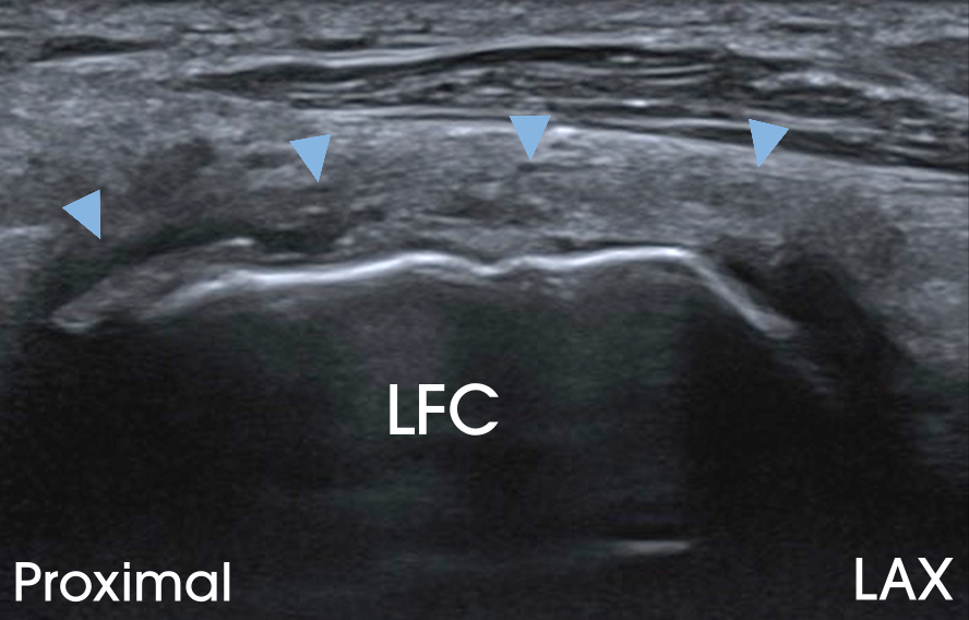
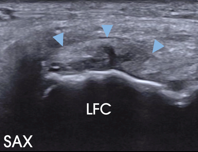
Ultrasound evaluation, including long and short-axis images, revealed a tear of the proximal lateral head of the gastrocnemius tendon (arrowheads) at its insertion onto the lateral femoral condyle (LFC).
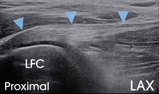
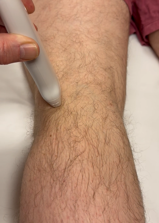
For reference, I’ve included a normal long-axis image of the lateral head of the gastrocnemius tendon (arrowheads), along with the corresponding probe position.
A subsequent MRI confirmed the diagnosis.
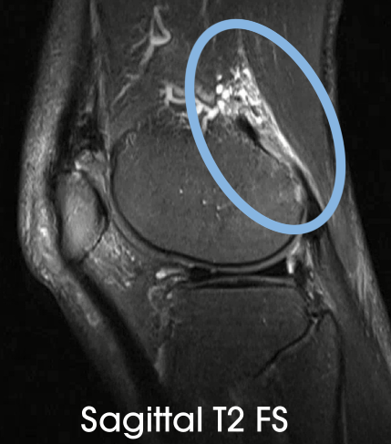
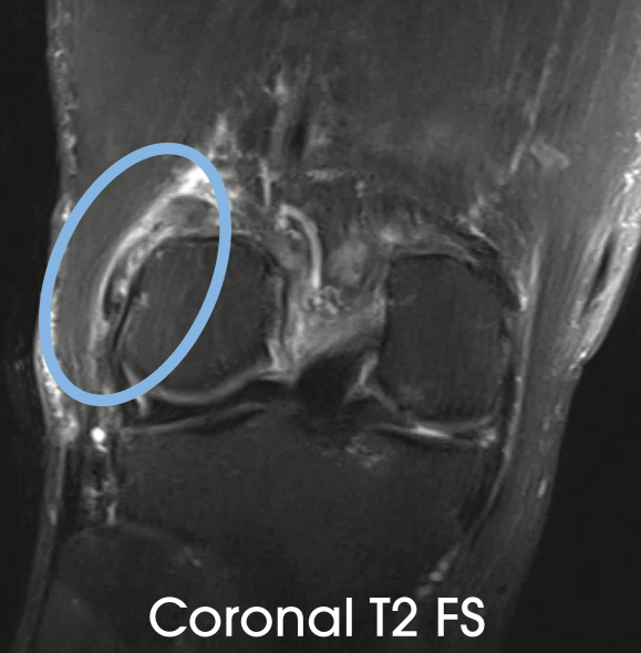
This case highlights the importance of evaluating the proximal tendon of the lateral head of the gastrocnemius when assessing posterolateral knee pain.
Thanks for this case. Just yesterday, I saw a patient with continued posterior lateral knee pain despite treatment for distal biceps femoris tendinopathy. I will keep the lateral gastroc in my differential moving forward as this is not something I thought much about yesterday. I ended up focusing more on the popliteus tendon origin which appeared abnormal compared to the non involved side.