
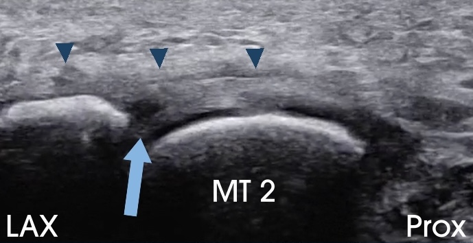
In the Forefoot Module, I present my scanning protocol and technique for evaluating the plantar plate of the lesser MTP joints. In this case, the long-axis (LAX) image of the plantar aspect of the 2nd MTP joint demonstrates a hypoechoic defect within the plantar plate (arrow), consistent with a tear. Note the fluid superficial to the proximal phalanx and within the plantar fat pad (arrowheads), findings that indicate a full-thickness tear with leakage of joint fluid into the plantar fat pad.
When assessing the plantar plate with ultrasound, it is important to include the short-axis (SAX) view. In this transducer orientation, the plantar plate should be followed to its enthesis on the medial and lateral condyles of the proximal phalanx. I pay particular attention to the lateral condyle, which lies in a more plantar position than the medial condyle and is therefore more vulnerable to repetitive loading and stress.
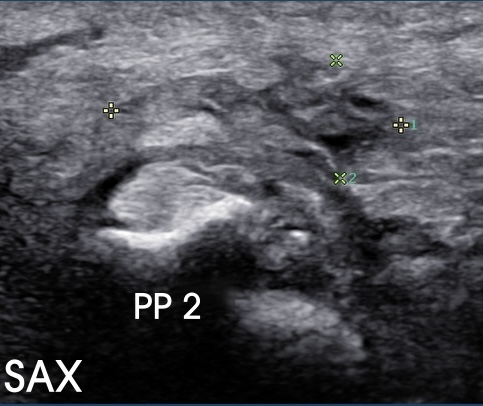
In this case, the SAX image shows bony irregularity of the lateral condyle of the proximal phalanx (ellipse). Such changes can serve as a direct pain generator and are often more clearly appreciated in the SAX than LAX. Importantly, in this patient, the bony changes at the lateral condyle corresponded to the point of maximal tenderness with sonopalpation.

A subsequent SAX image, obtained slightly distal, demonstrates hypoechoic thickening of the plantar fat pad superficial to the lateral condyle (between calipers). This finding further supports the role of the bony irregularity at the lateral condyle as a pain generator.
Summary: Always evaluate the plantar plate in both LAX and SAX planes. In SAX, image the plantar plate to its enthesis at the lateral condyle of the proximal phalanx, where pathology is common and may be underestimated on the LAX view. Fat pad abnormalities, especially when coupled with sonopalpatory pain, strengthen the case for bony changes of the lateral condyle as a significant pain generator.