
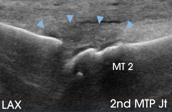
This 43-year-old active individual was referred for treatment of a presumed second metatarsal interspace neuroma. A diagnostic ultrasound was performed utilizing my forefoot scanning protocol, outlined in the Forefoot Module, which is designed to evaluate lesser metatarsalgia – pain arising from the 2nd through 5th metatarsophalangeal (MTP) joints and surrounding soft tissues. The protocol begins on the dorsal side of the foot with a long-axis (LAX) view of the MTP joints.
The patient’s plain radiographs demonstrated mild hallux valgus and early 1st MTP joint osteoarthosis. The LAX image below shows the 2nd MTP joint, where the joint capsule is enlarged (arrowheads).
When I detect a joint abnormality such as capsular enlargement, I make a point to also image the joint in the short-axis (SAX) plane, which can better demonstrate the extent of thickening. In this case, the SAX image below shows that the capsular thickening (arrowheads) extends into the 2nd metatarsal interspace (arrow), likely explaining the patient’s pain in that location. The subsequent ultrasound evaluation of the interspace itself was normal.

Take-home point: Always image MTP joint abnormalities in orthogonal planes to fully assess the extent of capsular thickening. A thickened capsule can encroach into the metatarsal interspace pain and mimic the symptoms of an interdigital neuroma.