
Acquired flatfoot deformity is often attributed to progressive dysfunction of the posterior tibialis tendon. However, medial ankle stability also depends on the integrity of the spring ligament and deltoid ligament complexes. When evaluating patients with acquired flatfoot deformity using ultrasound, I routinely assess the posterior tibialis tendon, the anteromedial band of the spring ligament, and the deltoid ligament complex.
In this case, a 74-year-old patient presented with a slowly progressive, minimally painful flatfoot deformity that developed over 10 years. The first video demonstrates a long-axis (LAX) view of the posterior tibialis tendon as it courses around the medial malleolus (MM). The tendon appears mildly thickened but remains intact. Note the hyperechoic aggregates within the tendon – a finding consistent with this patient’s long-standing history of gout.
The next video shows the anteromedial band of the spring ligament, which originates from the sustentaculum tali (ST) of the calcaneus, spans over the talar head, and inserts broadly onto the navicular (Nav). There is a focal defect just distal to the ST (blue arrow), consistent with a ligament tear. In the video, I gently evert and externally rotate the ankle to dynamically stress the ligament.
Next, I assess the deltoid ligament complex. The video below shows the tibiospring bundle of the deltoid ligament, which spans from the tibia (Tib) to the spring ligament (white arrow). The spring ligament appears hypoechoic due to a tear. A hypoechoic defect is seen where the fibers of the tibiospring bundle should insert onto the spring ligament.
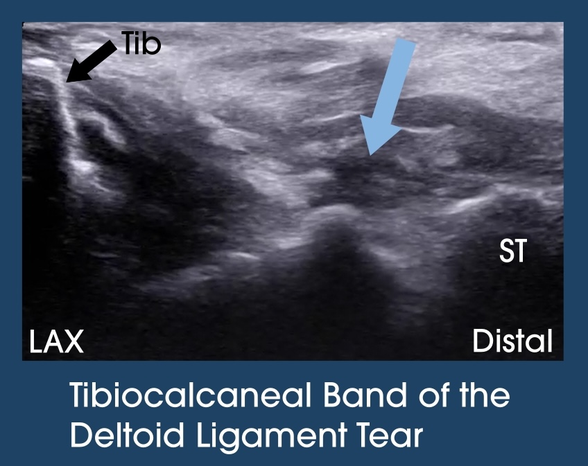
The final video is of the tibiocalcaneal bundle of the deltoid ligament, which spans from the tibia (Tib) to the sustentaculum tali (ST). The blue arrow highlights a hypoechoic defect in the ligament, consistent with a tear.
When assessing patients with acquired flatfoot deformity, I systematically evaluate the posterior tibialis tendon, anteromedial band of the spring ligament, and deltoid ligament complex.